SPATIAL FREQUENCY DOMAIN IMAGING (SFDI)
SFDI is a non-contact optical imaging method capable of quantitative imaging, spectroscopy, and tomography of tissues over a large field-of-view (many cm) and up to a depth of ~1cm. This method deals with imaging in the spatial frequency domain (SFD) and has also been referred to as modulated imaging (MI) in prior publications by this lab.
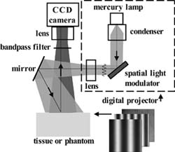
The imaging platform consists of three basic components: a light source, a spatial light modulator, and a CCD camera for detection (figure 1). A spatially-modulated illumination scheme with various spatial frequencies is projected over a large area of tissue and the remitted diffuse light is detected via a CCD camera and then demodulated in order to extract diffuse reflectance at multiple spatial frequencies. Figure 2 demonstrates this data flow for a measurement of vein structure in a human forearm. The diffuse reflectance at a certain frequency has differential sensitivity to absorption and scattering contrast in tissue. Due to this, the spatially-varying (2D or 3D) absorption and scattering characteristics of the sample can then be reconstructed from the measured diffuse reflectance using algorithms developed in the SFD.
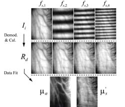
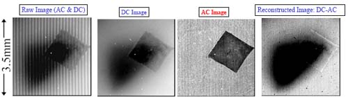
The absorption and scattering maps in figure 2 demonstrate vein structure. The estimation of intrinsic tissue chromophores is further enabled by quantifying absorption (with scattering correction) at many wavelengths. For example, images of tissue hemoglobin concentration (total, oxy- and deoxy-forms) and tissue oxygen saturation, lipid, water, and melanin concentration can all be quantified using appropriate red or infrared wavelengths. Similarily, this concept can be extended to fluoresence with appropriate spectral filtering. Finally, this technique has also demonstrated the ability for depth sectioning. Figure 3 demonstrates how higher spatial frequency (AC) is more sensitive to superficial absorbing stuctures (square) in turbid media than zero frequency (DC). Subsequently, simple image processing can be used to isolate a deeper structure (triangle).
Research in SFDI continues with the goal of developing novel tools for functional imaging. The research projects emphasize model development/validation in addition to in vitro and in vivo studies. These in vivo studies include research on traumatic brain injuries, melanoma, wound healing, tumor angiogenesis and chemotherapy among others.